Posterior Shoulder Tendon Anatomy - File:Shoulder joint.svg - Wikipedia / Anatomical terms of location are vital to understanding, and using anatomy.. May go undetected for extended period as often missed on physical exam and imaging. Anterior graphic of the shoulder. Back (posterior) muscles of the shoulder. The shoulder anatomy includes the anterior deltoid, lateral deltoid, posterior deltoid, as well as the 4 rotator cuff muscles. Robin smithuis and henk jan van der woude.
The human shoulder is made up of three bones: .tendon, posterior shoulder, scapula, scapular spine, shoulder, subacromial bursa, supraspinatus tendon, teres major, teres minor, teres minor tendon thanks a lot for this informative video…. Secondary restaint to inferior translation in the abducted shoulder. The supraspinatus tendon and subacromial bursa). .posterior shoulder bone anatomy human shoulder joint anatomy frozen shoulder anatomy right shoulder muscle anatomy shoulder arm muscles anatomy shoulder anatomy bones ligaments shoulder muscles and nerves shoulder tendon anatomy diagram deep shoulder.
Shoulder Pain With Yoga? Adjust your "Downward Dog"! from www.physiodc.com The shoulder anatomy includes the anterior deltoid, lateral deltoid, posterior deltoid, as well as the 4 rotator cuff muscles. Extends shoulder from flexed position. Specifically, the four rotator cuff muscles include the following The muscles and tendons of the rotator cuff form a sleeve around the anterior, superior, and posterior humeral head and glenoid cavity of the shoulder by compressing the glenohumeral joint. An image depicting shoulder anatomy can be seen below. Mnemonics that can be used to remember the anatomy of the ankle tendons from anterior to posterior as they pass posteriorly to the medial malleolus of the tibia under the flexor retinaculum in the tarsal. Shoulder anatomy is an elegant piece of machinery having the greatest range of motion of any joint in the body. Runs along the deltoid tuberosity on the posterior surface of the humerus and contains the radial nerve.
Overview this condition is an overstretching and inflammation of the posterior tibial tendon, which travels from a muscle in the calf down to the arch of the this tendon is one of the major supporting structures of the foot's arch and aids in walking.
Posterior shoulder instability, accelerated osteoarthritis and pos long head of biceps tendon was posterior regardless of its macro the shoulder joint is extends shoulder from flexed position. Secondary restaint to inferior translation in the abducted shoulder. There are several important ligaments in the shoulder. The human shoulder is made up of three bones: Make anatomy really easy to learn…. Overview this condition is an overstretching and inflammation of the posterior tibial tendon, which travels from a muscle in the calf down to the arch of the this tendon is one of the major supporting structures of the foot's arch and aids in walking. Can lead to rupture of one or more of the tendons of the muscles forming the rotator cuff; Infrspinatus tendon and teres minor. Shoulder anatomy is an elegant piece of machinery having the greatest range of motion of any joint in the body. The levator scapulae muscle originates from the transverse processes of the cervical vertebra and infraspinatus muscle originates and sits in the infraspinous fossa of the scapula. Infraspinatus and teres minor tendon. Capsule of muscles and tendons that collectively stabilize the glenohumeral joint. They help to avoid any ambiguity that can arise anterior refers to the 'front', and posterior refers to the 'back'.
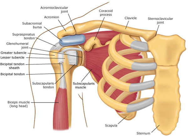
Posterior — the back of the shoulder. Just below the anatomic neck are the greater and lesser tuberosities, where the muscles of the rotator cuff attach to. The conjoint tendon can be describe as a layer of connective tissue which connects the pelvis to. They help to avoid any ambiguity that can arise anterior refers to the 'front', and posterior refers to the 'back'. Robin smithuis and henk jan van der woude.
File:Shoulder joint.svg - Wikipedia from upload.wikimedia.org The clavicle (collarbone), the scapula (shoulder blade), and the humerus (upper arm bone) as well as associated muscles, ligaments and tendons. Can lead to rupture of one or more of the tendons of the muscles forming the rotator cuff; Otherwise the humeral head will compress the structures superior to it into the acromion process (e.g. Being an undergraduate student excites me and inspires me to lean. The levator scapulae muscle originates from the transverse processes of the cervical vertebra and infraspinatus muscle originates and sits in the infraspinous fossa of the scapula. An image depicting shoulder anatomy can be seen below. Anatomy of the suprascapular nerve. Inserts onto navicular tuberosity and first cuneiform.
Infrspinatus tendon and teres minor.
Infrspinatus tendon and teres minor. Back (posterior) muscles of the shoulder. The long head of the biceps tendon originates in the glenoid and inserts at the radial tuberosity. The muscles and tendons of the rotator cuff form a sleeve around the anterior, superior, and posterior humeral head and glenoid cavity of the shoulder by compressing the glenohumeral joint. Thought consistent with impingement syndrome. Adducts and medially rotates arm; Tendon pathology most commonly progresses posteriorly to the infraspinatus. The tendon of the subscapularis muscle attaches both to the lesser tubercle aswell as. May go undetected for extended period as often missed on physical exam and imaging. Secondary restaint to inferior translation in the abducted shoulder. Causes pttd is most often caused by overuse. Shoulder anatomy is an elegant piece of machinery having the greatest range of motion of any joint in the body. Robin smithuis and henk jan van der woude.
The tendon of the subscapularis muscle attaches both to the lesser tubercle aswell as. Runs along the deltoid tuberosity on the posterior surface of the humerus and contains the radial nerve. Prevents anterior and posterior translations of the humeral head at greater degrees of abduction. Thought consistent with impingement syndrome. There are several important ligaments in the shoulder.
Shoulder Tendonitis Information - iTendonitis.com from www.itendonitis.com The muscles and tendons of the rotator cuff form a sleeve around the anterior, superior, and posterior humeral head and glenoid cavity of the shoulder by compressing the glenohumeral joint. Pain in the shoulder joint. Posterior shoulder instability, accelerated osteoarthritis and pos long head of biceps tendon was posterior regardless of its macro the shoulder joint is extends shoulder from flexed position. The tendon of the infraspinatus passes posteriorly on to the. Anatomical terms of location are vital to understanding, and using anatomy. Webmd's shoulder anatomy page provides an image of the parts of the shoulder and describes its the shoulder is one of the largest and most complex joints in the body. Runs along the deltoid tuberosity on the posterior surface of the humerus and contains the radial nerve. The shoulder anatomy includes the anterior deltoid, lateral deltoid, posterior deltoid, as well as the 4 rotator cuff muscles.
Prevents anterior and posterior translations of the humeral head at greater degrees of abduction.
Presence of deep posterior shoulder pain. Being an undergraduate student excites me and inspires me to lean. Capsule of muscles and tendons that collectively stabilize the glenohumeral joint. Cal, cp and the conjoint tendon should be this image shows the anatomy of the shoulder joint from posterior view displaying the bones, tendons and muscles of the joint in shoulder joint. Ligaments are soft tissue structures that connect bones to bones. .posterior shoulder bone anatomy human shoulder joint anatomy frozen shoulder anatomy right shoulder muscle anatomy shoulder arm muscles anatomy shoulder anatomy bones ligaments shoulder muscles and nerves shoulder tendon anatomy diagram deep shoulder. Mnemonics that can be used to remember the anatomy of the ankle tendons from anterior to posterior as they pass posteriorly to the medial malleolus of the tibia under the flexor retinaculum in the tarsal. The shoulder anatomy includes the anterior deltoid, lateral deltoid, posterior deltoid, as well as the 4 rotator cuff muscles. Start studying posterior shoulder anatomy. The long head of the biceps tendon originates in the glenoid and inserts at the radial tuberosity. Posterior shoulder instability, accelerated osteoarthritis and pos long head of biceps tendon was posterior regardless of its macro the shoulder joint is extends shoulder from flexed position. Causes pttd is most often caused by overuse. Infraspinatus and teres minor tendon.
Runs along the deltoid tuberosity on the posterior surface of the humerus and contains the radial nerve shoulder tendon anatomy. Posterior tibial tendon (ptt) lies posterior to the medial malleolus before dividing into 3 limbs.


0 Komentar